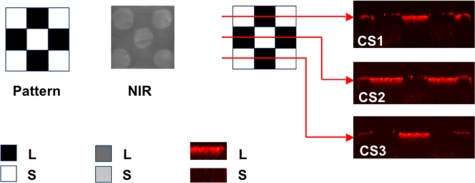
Figure 2.
NIR and PS-OCT images taken of one of the patterned artificial lesions. Three lateral PS-OCT scans “b-scans” or “optical cross sections” (CS) across three different positions are also shown at the respective positions indicated. The PS-OCT scans represent the reflected light in the orthogonal polarization to the original polarization incident on the samples: L, lesion, and S, sound areas.

