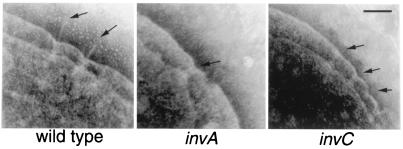
Figure 1.
Role of the type III secretion protein-export apparatus in the assembly of the S. typhimurium needle complex. Electron micrographs of negatively stained, osmotically shocked wild-type S. typhimurium and the isogenic invA and invC mutant derivatives. The positions of needle complexes and bases are indicated by arrows. (Bar = 100 nm.)

