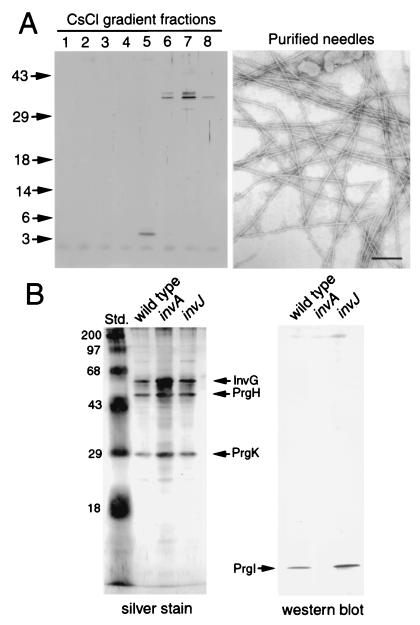
Figure 4.
Isolation and identification of the main subunit of the needle substructure. (A) Needle structures were isolated from a S. typhimurium invJ mutant strain and purified on a CsCl density gradient, and the different fractions were loaded on a SDS/polyacrylamide gel and visualized by silver staining. An electron micrograph of negatively stained purified needle structures from fraction 5 of the CsCl gradient is shown. (Bar = 100 nm.) (B) Purified needle complexes from wild-type S. typhimurium and the isogenic invA or invJ mutants were separated on a SDS/polyacrylamide gel and visualized by silver staining or transferred to nitrocellulose membranes and probed with an antibody directed against PrgI. The identities of the different proteins are indicated.

