
Figure 2.
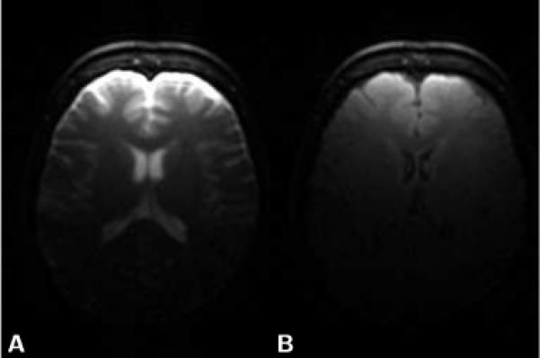
Axial T2-weighted (b=5 s/mm2) brain images obtained with LSDI. The image shown in A) was obtained without fluid signal suppression. For the image shown in B) fluid signal suppression was applied with a 2400 ms delay between magnetization inversion and column scanning. Other imaging parameters remained unchanged.