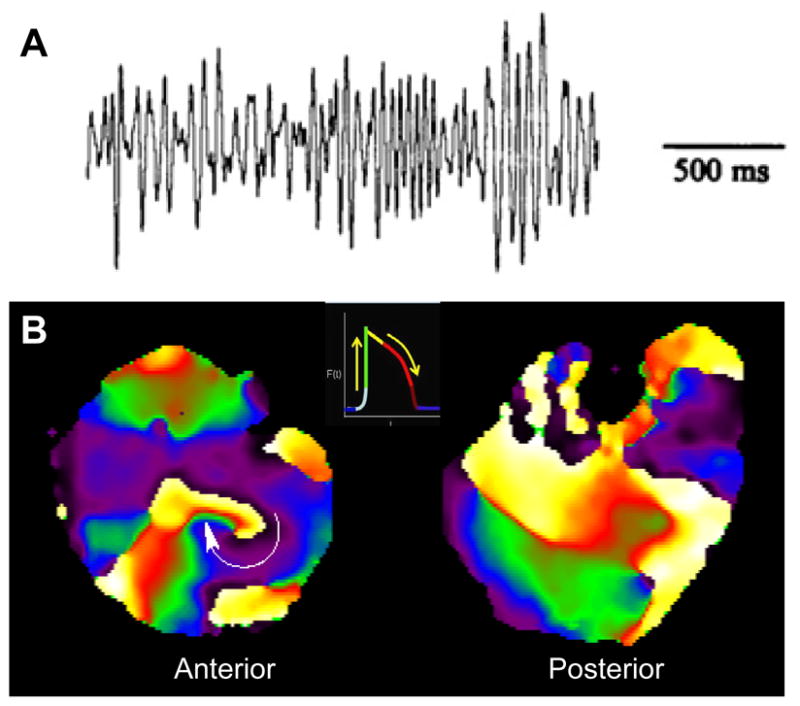
Figure 2.

High-frequency stationary rotor results in fibrillation in an isolated, Langendorff-perfused guinea pig heart. A, ECG trace of VF. B, Snapshots of phase maps of the anterior (left) and posterior (right) epicardial surfaces of the ventricles during VF, which was maintained by a long-lasting rotor rotating clockwise on the anterior left ventricular wall. Colors indicate different phases of the action potential (inset) at each pixel location.
