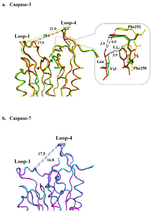
Fig. 8.
Comparison of caspase complexes with pentapeptides and tetrapeptide. (a) Superposition of Cα backbone of caspase-3 with LDESD (red), VDVAD (green) and DEVD (yellow). Interatomic distances in Å are shown by broken lines. Conformational change is indicated by the altered separation of loop-1 and loop-4 in different complexes. The S5 binding site (boxed region) is shown in detail, where Phe250 and Phe252 form hydrophobic interactions with the P5 residues. (b) Superposition of Cα backbone of caspase-7 with LDESD (cyan) and DEVD (magenta). No significant structural change is observed in the two complexes of caspase-7.

