
Figure 2. Tt-catl and Tt-cysb mRNA location by in situ hybridization.
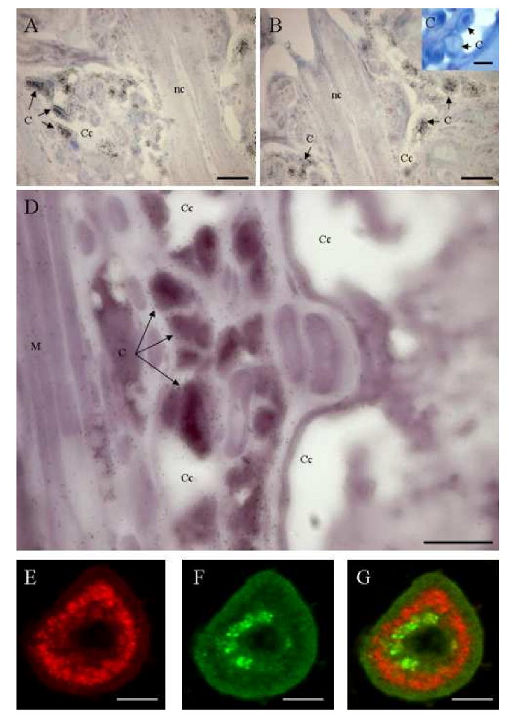
A, B, C. Detection of Tt-catl mRNA in leech tissues by in situ hybridization. Paraffin-embedded sections of animals were hybridized with antisens (A, B) Tt-catl riboprobes were labelled with 35S-UTP. Positive circulating cells are detected in cœlomic cavities and in the ventral sinus. Negative controls consisting of sections hybridized with Tt-catl sense riboprobes (C). D. Double detection of Tt-catl and cystatin b mRNAs (Tt-cysb). Sections were hybridized with antisens Tt-catl riboprobes labelled with Dig-UTP and with antisens Tt-cysb labelled with 35S-UTP. Both probes are co-localized in cœlomocytes. E, F. Confocal microscopic images of Tt-CATL (red) and Tt-CYSB (green) double immune labelling in leech cœlomocytes. G is the merged confocal image of E and F. Merged confocal image suggests that Tt-CATL and Tt-CYSB may be packed in the same cells but in different cell compartments. Abbreviations: C. Cœlomocytes. Cc. Cœlomic cavity. Nc. Nerve cord. M. Muscle. Bars: A, B, C and D: 100 μm. E, F and G: 30 μm.