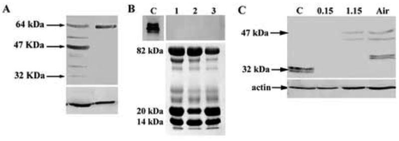
Figure 3.

(A) Western blotting of cell lysates obtained by epithelial scrape from human corneal epithelium and limbus for IGFBP3 using a goat polyclonal antibody. Blot representative of repeated experiments (n=2). (B) Western blot for IGFBP3 from basal, resting tear samples. IGFBP3 was not detectable in human tear samples (collected from 3 independent patients, represented 1, 2, and 3, top). rhIGFPB3 served as a positive control, C. Sypro orange staining confirmed presence of protein in tear samples (bottom). (C) Western blotting for IGFBP3 in hTCEpi cells grown in submersed/airlifted culture. IGFBP3 protein was undetectable in hTCEpi cells under 0.15 mM calcium culture conditions. Appearance of IGFBP3 isoforms correlated with an increase in calcium (1.15 mM) levels and subsequent air-lifted (Air) differentiation. B-actin was used as a loading control (n=2).
