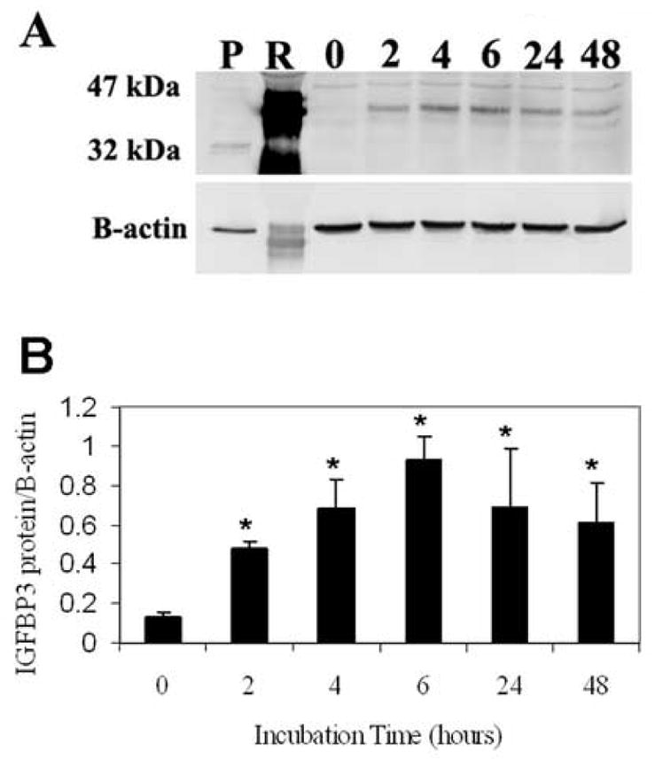
Figure 6.

(A) hTCEpi cells grown on plastic and incubated with 20 μg of rhIGFBP3 demonstrated a corresponding increase in IGFBP3 protein levels. B-actin was used as a loading control. P: IGFBP3 plasmid expressed in hTCEpi cells as a positive control; R: 20 μg of recombinant human protein loaded into lane 2 as a positive control; 0 – 48 represent incubation times in hours. (B) Quantitative analysis of IGFBP3 as a function of incubation time. The addition of rhIGFBP3 resulted in an increase in cellular associated IGFBP3 compared to the control (n=3, mean ± standard deviation of 3 repeated experiments, One-way ANOVA, P<0.001).
