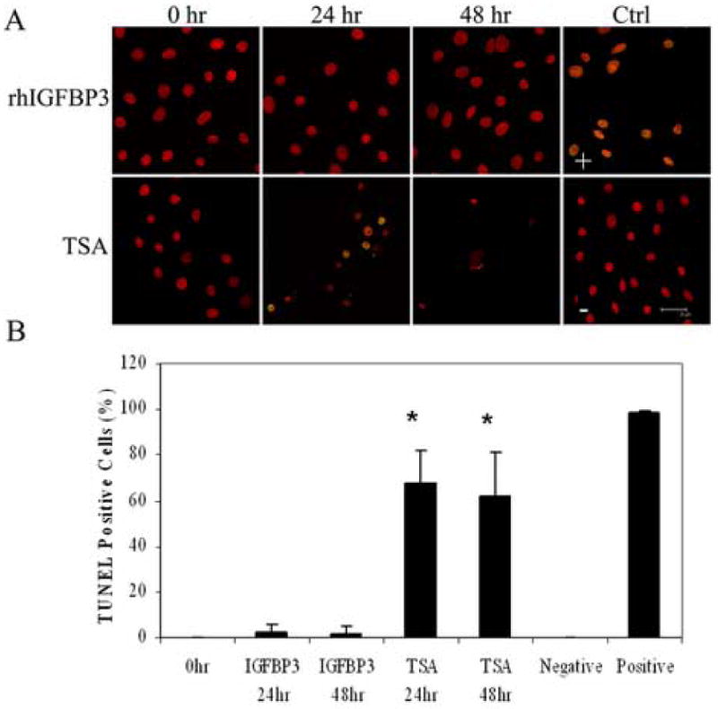
Figure 9.

(A) Double-labeling of hTCEpi cells with TUNEL (green) and PI (red) on collagen-coated glass coverslips and treated with 500 ng/ml rhIGFBP3 or 3.31 μM TSA for 0, 24, and 48 hours. Positive control (+): cells treated with DNase; negative control (-): TdT Enzyme omitted. Scale: 28 μm. (B) Percentage of TUNEL positive cells following treatment with TSA and rhIGFBP3 protein. TSA induced significant cell death (n=2, graph representative of repeated experiments, One-way ANOVA, P<0.001). At 48 hours, robust cell loss was seen due to apoptosis, making quantification difficult. 63× magnification, scale: 28 μm.
