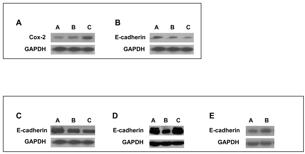
Figure 1. IL-1β -dependent regulation of COX-2 and E-cadherin in HNSCC.
IL-1β regulates expression of COX-2 and E-cadherin in a dose dependent manner. Tu686 cells were treated with the indicated concentrations of IL-1β for 18 h. Protein from whole cell lysates of was analyzed for COX-2 and E-cadherin expression by Western Blot as described in Materials and Methods. (A) IL-1β up-regulates COX-2 expression in a concentration-dependent manner. Lane A: no treatment; Lane B: IL-1β (100 U/ml); Lane C: IL-1β (200 U/ml). (B) IL-1β causes down-regulation of E-cadherin expression in a concentration-dependent manner. Lane A: no treatment; Lane B: IL-1β (100 U/ml); Lane C: IL-1β (200 U/ml). (C) Upon addition of celecoxib, E-cadherin is no longer down-regulated in Tu686 HNSCC cells indicating that functional COX-2/PGE2 is required for its down regulation. Lane A: IL-1β (100 U/ml) + celecoxib (1µM); Lane B: diluent (0.1% BSA in 1x PBS) alone; Lane C: IL-1β (100 U/ml). (D) In the presence of COX-2 shRNA, E-cadherin is no longer down-regulated in Tu686 HNSCC cells indicating that functional COX-2/PGE2 is required for its down regulation. Lane A: diluent (0.1% BSA in 1x PBS) alone; Lane B: IL-1β (100 U/ml); Lane C: IL-1β (100 U/ml) + COX-2 shRNA. (E) PGE2 causes the down-regulation of E-cadherin expression in Tu686 cell lines. Lane A: PGE2 (10µg/ml); Lane B: diluent (0.1% BSA in 1x PBS) alone.

