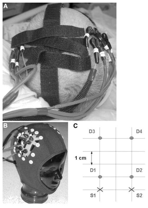
Fig. 1.
NIRS probes are attached to a patient’s head at the bedside using Velcro® strips (a) or a neoprene skull cap (b). c The source-detector geometry overlying each hemisphere consists of two source positions (S1, 2) two cm apart, and four detector positions (D1–4) at one and four cm distance from each source

