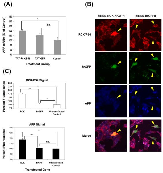
Figure 6.
Rck/p54 overexpression increases APP mRNA and protein levels in SH-SY5Y cells. (A) 50nM TAT-RCK or TAT-GFP was transduced into SH-SY5Y cells. APP mRNA levels were quantified by real time PCR. The amount of signal, n = 3, was quantified and normalized to the amount present in untreated control cells, ± S.E.M. *: p<0.05; N.S. = not significant (p > 0.1) (two-tailed, homoscedasic T-test). (B) SH-SY5Y cells were transfected with various expression vectors as shown. Eighteen hours later, cells were fixed and stained with rhodamine-conjugated anti-rck/p54 and AlexaFluor® 633 conjugated anti-APP antibodies. Representative positive transfected cells are shown. (C) APP and rck/p54 staining was quantified for cells transfected with pIRES-RCK-hrGFP II (RCK) or pIRES-hrGFP II (hrGFP). The amount of signal from approximately 50 positively transfected cells was quantified and normalized to approximately the same number of untransfected cells. (*: p<0.05; ***: p <0.001; N.S. = not significant (p > 0.1) (two-tailed, heteroscedasic T-test).

