
Figure 5.
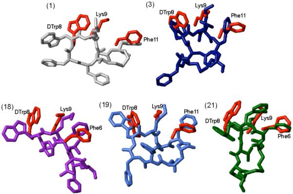
Superposition of sst4 receptor-specific pharmacophore with the 3D NMR structures of the analogues 1, 3, 18, 19 and 21. The sst4 pharmacophore34 is represented by amino acid side chains colored in red. The conformer with the lowest energy represents the 3D structures of the analogues and they are color coded as in Figure 2. In addition, for each analogue the amino acid side chains proposed to be involved in receptor binding are labeled.