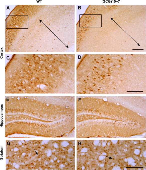
Figure 6.

Calbindin D28K-positive interneurons are less abundant in the neocortex, hippocampus, and striatum of the Arx(GCG)10+7 mutant. A, B, Neocortex of WT and mutant motor/somatosensory cortex immunostained with anti-calbindin D28K antibody, showing loss of calbindin+ cells throughout the Arx(GCG)10+7 cortex compared with a WT sibling. Layers V–VI are indicated by the two-headed arrows. C, D, Higher magnification images (A, B, rectangles) show calbindin+ interneurons in layers I–IV. E, F, Calbindin+ interneurons are also decreased in the granule cell layer of the mutant dentate gyrus. G, H, Like the ARX+ interneurons, calbindin+ projection neurons (arrows) in the mutant striatum are reduced to approximately one-half the WT level. Scale bars: A, B, E, F, 200 μm; C, D, G, H, 100 μm.
