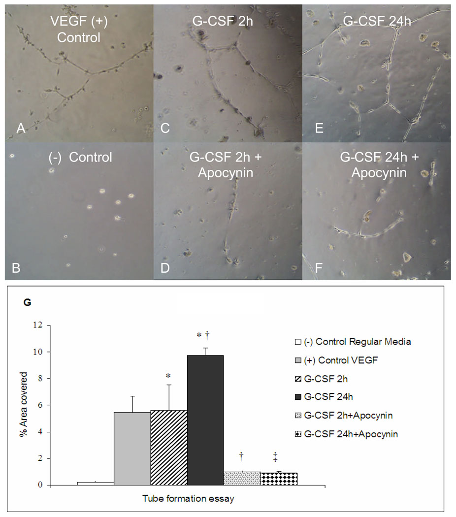
Figure 6.
Human coronary artery endothelial cell (HCAEC) tube formation in Matrigel in response to VEGF or to conditioned media from cardiac myocytes treated with G-CSF for 2 or 24 hrs. A: percentage of area covered with tubes (n=4/group, means±SE). * vs VEGF, † vs G-CSF 2h, ‡ vs G-CSF 24h, P <0.01). B–G: Images of tube formation after addition of G-CSF cardiomyocyte stimulation medium and apocynin (300 µM), as indicated.

