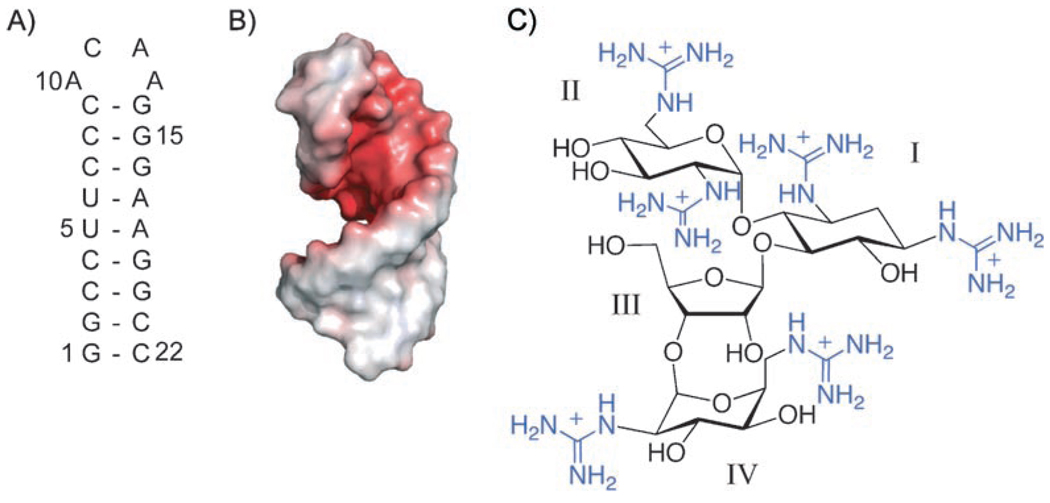
Figure 1.
HIV-1 frameshift site and guanidinoneomycin B. A) Sequence and secondary structure of the stem-loop RNA of the HIV-1 frameshift site. The numbering for the stem-loop RNA construct begins with the 5′ guanosine residue as 1. B) Electrostatic surface potential of the frameshift-site stem loop. Electrostatic potential is indicated by color (red = −4 kT/e, white = 0 kT/e, blue = +4 kT/e). C) Chemical structure of guanidinoneomycin B. The numbering of the individual glycosidic rings is indicated with Roman numerals. Guanidinium groups are in blue.

