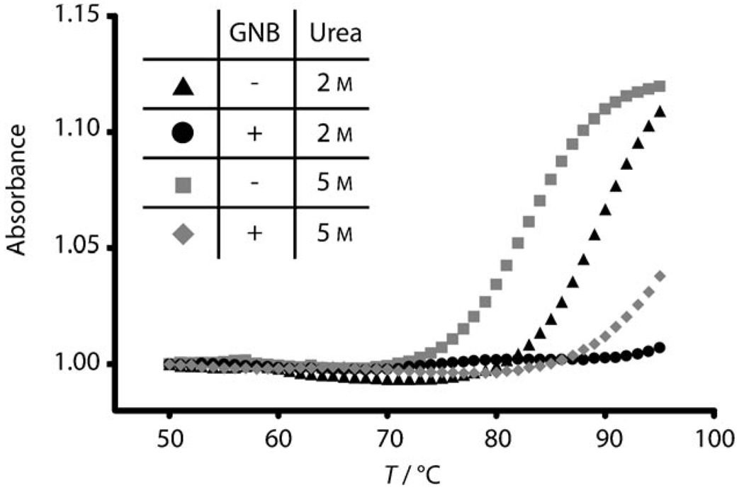
Figure 2.
Thermodynamic denaturation profiles of the HIV-1 frameshift-site RNA with and without GNB at varying concentrations of urea. UV absorbance at 260 nm is plotted versus temperature for the frameshift-site stem-loop RNA (2 µm) in the absence of GNB with urea at a concentration of 2 m (▲) or 5 m ( ). The sample of the GNB-bound RNA contained RNA (2 µm) in the presence of GNB (2.5 µm) with urea at a concentration of 2m (●) or 5 m (
 ). The RNA–GNB complex was formed at high concentration (1.2 mm RNA+1.5 mm GNB) and diluted immediately prior to analysis. The low-temperature baseline absorbance was normalized to 1 for all samples.

