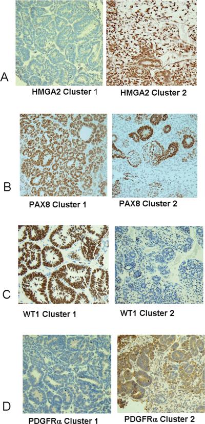
Figure 3. Immunohistochemistochemical validation of protein expression in VLRWT.
A. Immunohistochemical analysis for HMGA2: All four cluster 1 tumors were negative (left); all seven tumors within clusters 2 were immunoreactive for HMGA2 in greater than 10% of the cells.
B. Immunohistochemical analysis for PDGFRa: All four cluster 1 tumors showed immunoreactivity in fewer than 10% of cells (left); of seven cluster 2 tumors, fewer than 10% of the cells were immunoreactive in three tumors, and greater than 10% of the cells were immunoreactive in four tumors (right).
C. Immunohistochemical analysis for WT-1: All four cluster 1 tumors were immunoreactive for WT1 in greater than 10% of the cells (left); of seven cluster 2 tumors, five showed fewer than 10% of the cells to be positive and two showed greater than 10% of the cells positive (right).
D. Immunohistochemical analysis for PAX8: All four cluster 1 tumors showed greater than 80% of the cells to be immunoreactive for PAX8; of six evaluable cluster 2 tumors, all showed greater than 10% of the cells to be immunoreactive (right).

