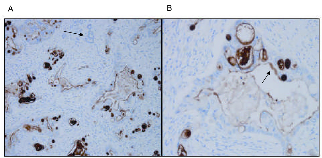
Figure 2. Human pancreatic adenocarcinoma expresses mesothelin.
Representative micrographs at low (A) and high (B) power of human pancreatic adenocarcinoma stained for mesothelin. Tissue sections were stained with the anti-human mesothelin mAb, 5B2 and counterstained by H&E. Non-tumor pancreatic glands are nonreactive with the mesothelin antibody, as shown by the arrow in panel A. Only tumor glands (see arrow in B) stain positive for mesothelin. The malignant tumor epithelium is 3+/3 positive in all the tumor glands.

