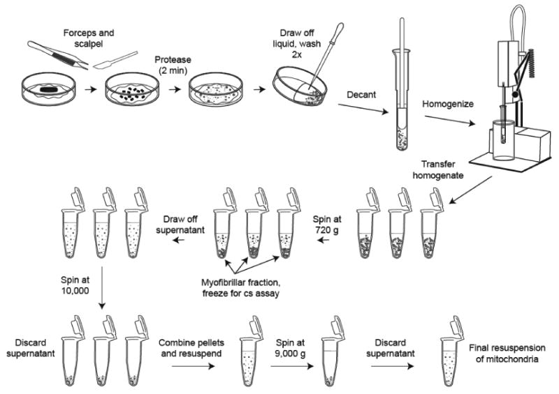
Figure 2. Tissue homogenization and isolation of mitochondria.

40-100 mg of muscle tissue is placed on an ice-cooled Petri dish and finely minced with forceps and scalpel. A 2-minute protease incubation is used to soften the tissue to help liberate mitochondria that are tightly bound to the contractile filaments. The protease is removed by diluting and washing with buffer. An all glass Potter-Elvehjem tissue grinder (0.3 mm clearance) is used to gently homogenize the tissue in an ice bath. The mitochondria are isolated by differential centrifugation. A low speed spin first removed the myofibrillar portion. A series of two high speed spins generates a mitochondrial pellet, which is resuspended in buffer for functional measurements.
