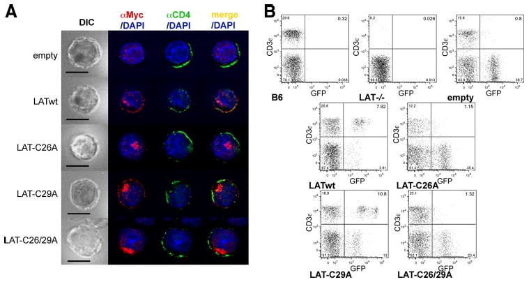
Figure 3.

Palmitoylation of LAT Cys26 is sufficient for LAT PM localization and T cell development. A, Mouse CD4+ T cells were infected with empty vector, LATwt, LAT-C26A, LAT-C29A, or LAT-C26/29A, stained with anti-Myc to localize exogenous LAT variants (red), with anti-CD4 (green), and with DAPI (blue), and then analyzed by fluorescence microscopy. One representative cell from at least 25 cells analyzed per group is shown. Bar, 10 μm. B, Lat−/− BM cells were infected as in A and then transferred into irradiated B6 mice. After 6 wk blood was analyzed for the presence of T cells. Untreated B6 and Lat−/− mice were used as controls. Diagrams are representative for one of four mice per group from two independent experiments.
