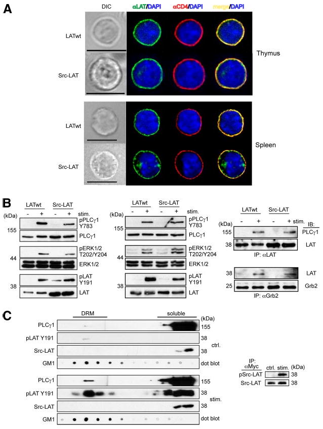
Figure 6.

Src-LAT reconstitutes TCR signaling in Lat−/− BM chimeric mice. A, Thymocytes and splenocytes from Lat−/− BM chimeric mice were stained with anti-LAT (green), anti-CD4 (red), and DAPI (blue) and then analyzed by fluorescence microscopy. Depicted images are representative of at least 20 GFP+ cells each. Bar, 10 μm. B, Thymocytes (left panel) and splenic CD4+ T cells (middle and right panels) from Lat−/− BM chimeric mice were stimulated for 1 min with anti-CD3 plus anti-CD4 mAbs, or for 2 min with anti-CD3ε and anti-CD28 mAbs, respectively. Cell lysates were directly immunoblotted (left and middle panels) or first immunoprecipitated with anti-LAT (upper right panel) or anti-Grb2 (lower right panel). Representative blots from two independent experiments are shown. C, Src-LAT infected CD4+ T cells from B6 mice were left unstimulated (upper left panels) or stimulated with anti-CD3ε plus anti-CD28 mAbs for 2 min (lower left panels), lysed, and then subjected to sucrose gradient fractionation. Fractions were immunoblotted with cholera-toxin B subunit to identify DRM fractions (GM1), with anti-PLCγ1, with anti-Myc to detect exogenous Src-LAT, and with anti-phospho LAT Y191 (pLAT Y191) to label phosphorylated endogenous LAT (wt) and exogenous Src-LAT (indistinguishable by size). Src-LAT was immunoprecipitated from the soluble fraction with anti-Myc and then blotted with anti-pTyr and anti-LAT Abs (right panel). Representative blots from two independent experiments are shown.
