
Figure 1. Identification of a 44-bp Mef2c endothelial-specific enhancer.
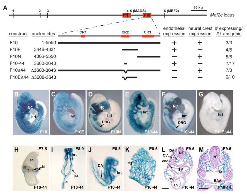
(A) A schematic representation of the mouse Mef2c locus is shown on the top line with exons depicted as vertical lines. The red boxes denotes the sizes and positions of the F7 and F10 fragments. F10 contains three evolutionarily conserved regions, denoted CR1-3. The lower portion of (A) depicts the deletion constructs of Mef2c F10. CR3 contains a neural crest specific enhancer. CR2 contains an endothelial specific enhancer, which encompasses a 44-bp deeply conserved region that is sufficient for endothelial enhancer activity in vivo. Endothelial and neural crest activity of each of the deletion constructs is denoted at the right as a + or -. The total number of transgenic embryos and the number that directed β-galactosidase expression to either the neural crest or endothelium are denoted at the far right of (A).
(B-G) Representative X-gal stained transgenic embryos for each of the Mef2c F10 transgene deletion constructs depicted in (A).
(H-M) Expression of the Mef2c F10-44-lacZ construct is specific to endothelial cells from blood island (bl) stage at E7.5 (H) throughout early endothelial development at E8.0 (I) and E8.5 (J, K). Transverse sections through an X-gal stained E9.5 transgenic embryo (L, M) demonstrate that transgene expression is restricted to endothelial cells throughout the vasculature, including the endocardium (end). al, allantois; BAA, branchial arch artery; CV, cardinal vein; DA, dorsal aorta; DRG, dorsal root ganglia; ec, ectoplacental cone; hrt, heart; LV, left ventricle; NC, neural crest; NT, neural tube; RV, right ventricle; SV, sinus venosus; YS, yolk sac.