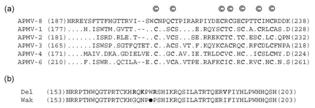
Fig. 3.
The V and W proteins. (a) Sequence alignment of the C-terminal end of V proteins of different APMVs (‘©’ denote the conserved cysteine residues that are also given in bold letters; the numbers indicate amino acid positions). (b) Sequence alignment of the C-terminal ends of the putative W protein of APMV-8 strains Delaware (Del) and Wakuya (Wak) (the opal codon (UGA) of W protein in Wakuya strain (●) terminates the reading frame after 172 aa. The amino acid differences between the strains are given in bold and underlined).

