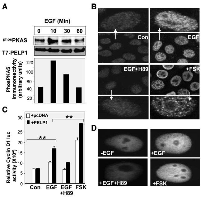
Figure 2.
Growth factors promote phosphorylation of PELP1 via PKA. A. MCF-7-PELP1 cells were serum starved for 24 h and stimulated with EGF (100 ng/mL) for the indicated periods of time, and T7-PELP1 was immunoprecipitated. The status of PKA-mediated phosphorylation on PELP1 was analyzed by using the PKA antibody. B. MCF-7 cells were treated with EGF in the presence or absence of the PKA inhibitor H89, and the cellular localization of PELP1 was determined by using confocal microscopy. C. Cos1 cells were transfected with the cyclin D1 luciferase reporter, along with pcDNA vector or pcDNA-PELP1. Cells were serum starved for 24 h, treated with EGF, EGF + H89, or forskolin for 12 h. The luciferase activity was then measured. *, P < 0.05; **, P < 0.001. Columns, mean from three independent experiments done in triplicate wells; bars, SE. D. Cos1 cells were transfected with GFP-PELP1-WT vector and treated with EGF (100 ng/mL) in the presence or absence of the PKA inhibitor H89, and the cellular localization of GFP-PELP1 was determined by using confocal microscopy.

