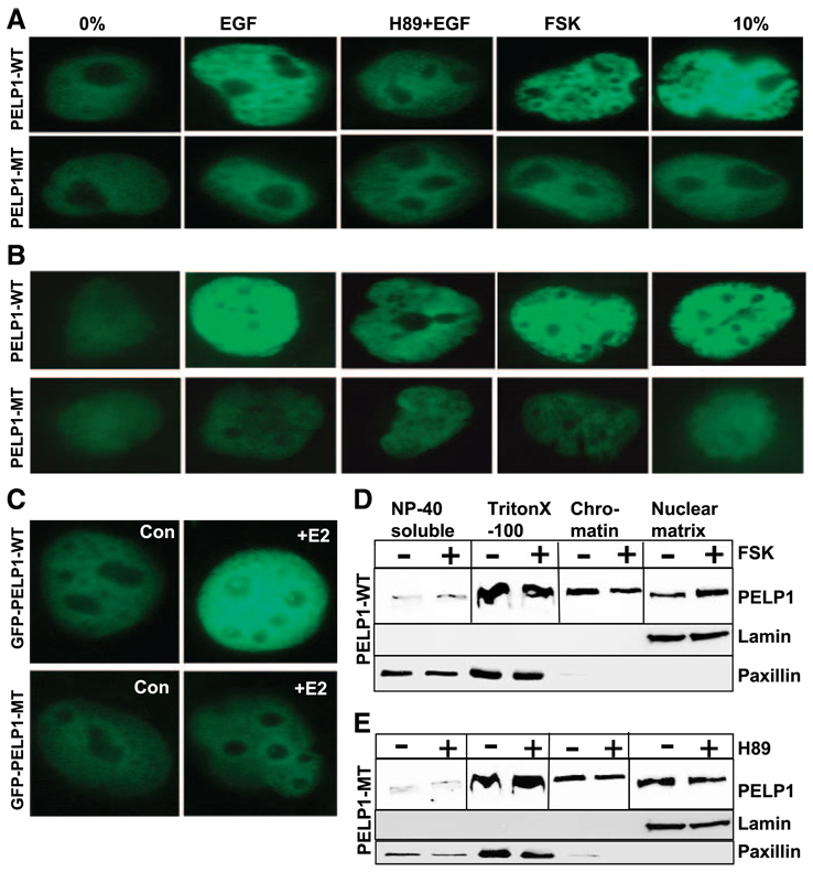
Figure 5.
PKA promotes nuclear redistribution of PELP1. MCF-7 cells (A) or T47D cells (B) were transfected with GFP-PELP1-WT or GFP-PEFP1-MT (S350, S415, S613A). After 48 h, cells were serum starved for a further 24 h, stimulated with serum, EGF, EGF + H89, or forskolin. Localization of GFPPELP1-WT and GFP-PELP1-MT (S350, S415, S613A) was determined by using confocal microscopy. C. MCF-7 cells were transfected with GFP-PELP1-WT and GFP-PELP1-MT (S350, 415, 613A) and treated with or without E2. The cellular localization of GFP-PELP1 was determined by using confocal microscopy. D and E. MCF-7cells were serum starved, treated with either forskolin (D) or H89 (E), and serially fractionated into NP40-soluble, cyto/ nucleoplasmic, chromatin, and nuclear matrix fraction. Equal amounts of protein were analyzed for localization of PELP1 by using Western blot analysis.

