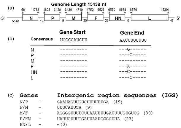
Fig. 2.
APMV-9 gene order, putative transcription signals, and IGS. (a) Schematic representation of the APMV-9 genome with the nt coordinates of the genes shown above the line and the lengths of the 3′ leader, 5′ trailer, and IGS shown below the line. (b) APMV-9 gene-start and gene-end sequence motifs. (c) APMV-9 IGS. All sequences are in negative-sense.

