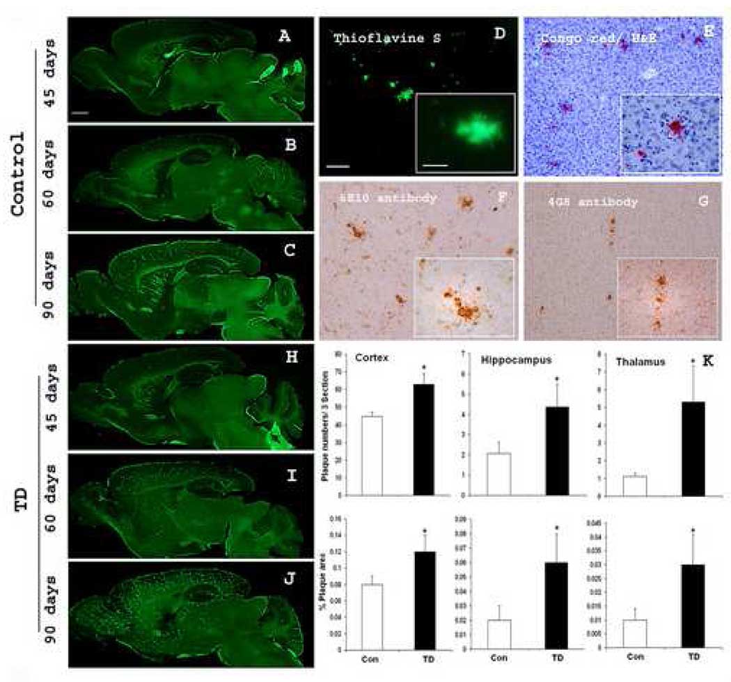
Fig. 4. TD accelerates deposition of amyloid plaques.
A–C show representative sagittal brain sections from 45, 60 and 90 days old Tg19959 mice stained with thioflavine S. D–G illustrates the profiles of amyloid plaques by different staining methods or antibodies: thioflavine S (D) and congo red (E), 6E10 antibody (F) and 4G8 antibody (G). H–J show representative sagittal brain sections from 45, 60 and 90 days old Tg19959 mice made TD for 10 days stained with thioflavine S. (K) shows the plaque counts and % area occupied by plaques quantified from the cortex, hippocampus and thalamus region. A statistical comparison of the sexes within groups did not find any sex differences. In controls, we compared males and females and found no differences (p>0.05). In TD we compared males and females and found no differences (p>0.05). The statistical comparisons were done both by one way ANOVA and Student t test. Data represent means ± SEM; Control (n=9) and TD (n=10) from 2–3 independent experiments. Scale bars: A–C and H–J, 500 µm; D–G, 100 µm; Insets 20 µm.

