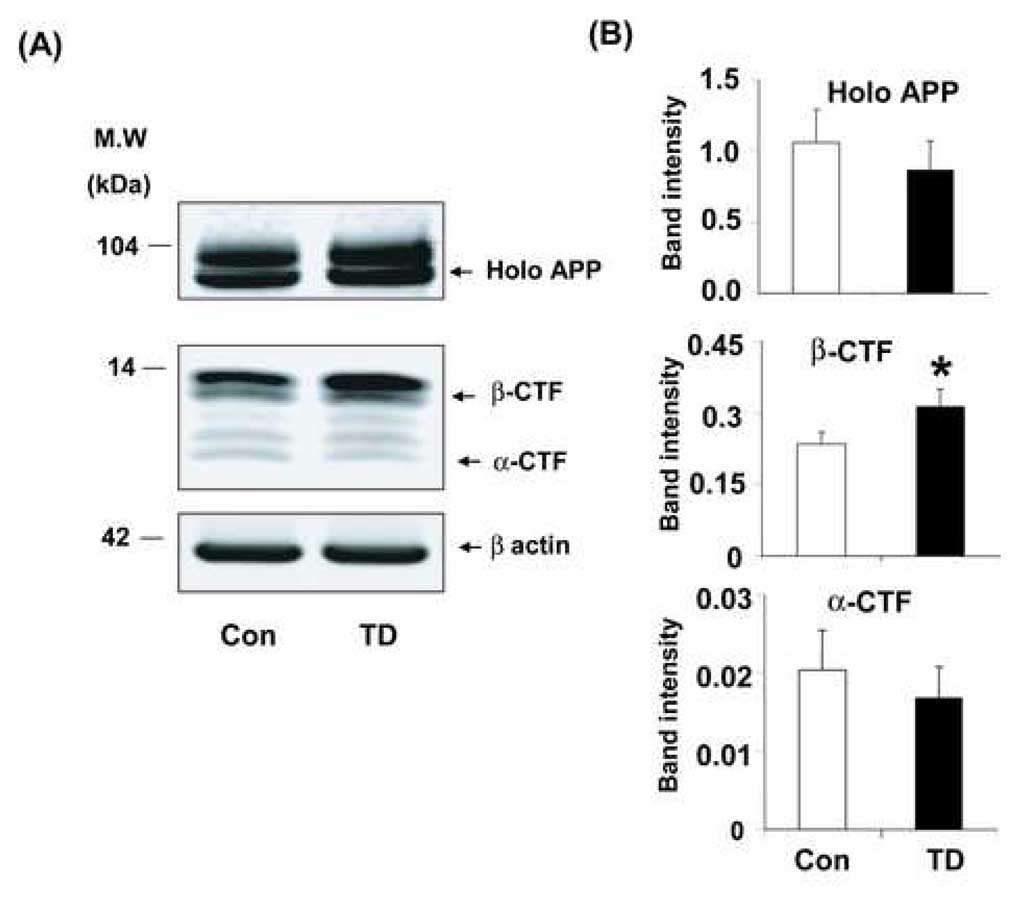
Fig. 6. TD increased APP C-terminal fragments in Tg19959 mice.
Sixty days old Tg19959 mice that were made TD for 10 days were compared to appropriate control animals. Panel (A) shows representative Western blots probed with G369 antibody, an antibody specific against both APP holo protein and C-terminal fragments. Panel (B) shows the densitometric analysis of APP holo protein and C-terminal fragments band intensity. The values represent the results from three independent experiments. Data represent mean ± SEM; Control (n=9) and TD (n=8). * p<0.05, TD vs control.

