Figure 2.
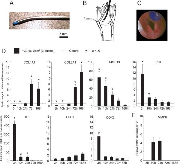
In vivo experimental procedures and real time RT-PCR analysis showing expression of select ECM and inflammatory cytokine genes in PDL-treated vocal folds. A: Angled stainless-steel rigid catheter allowing passage and directional control of the PDL fiber optic cable under endoscopic guidance. B: Schematic showing the catheter positioned with the fiber tip 1 mm above the membranous vocal fold surface. C: Endoscopic image showing the fiber tip positioned 1 mm above the membranous vocal fold surface prior to tissue irradiation. D: Fold change in relative mRNA expression of COL1A1, COL3A1, MMP13, IL1B, IL6, TGFB1 and COX2 in PDL-treated vocal folds compared to control, 3–168 h following delivery of ~39.46 J/cm2 laser fluence. E: Relative mRNA expression of MMP8 in PDL-treated vocal folds, 3–168 h following delivery of ~39.46 J/cm2 laser fluence. MMP8 was not detected in control, 3 h, 72 h and 168 h samples. Data represent four animals per time point. ACTB was employed as a housekeeping gene.

