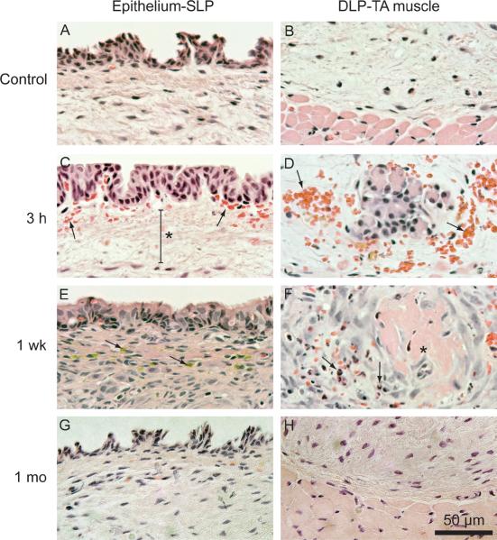
Figure 3.
Representative hematoxylin-eosin stained vocal fold sections of tissue harvested 3 h, 1 week and 1 month following delivery of ~39.46 J/cm2 laser fluence. A: Control section showing the epithelium and superficial lamina propria (SLP). B: Control section showing the deep lamina propria (DLP) and thyroarytenoid (TA) muscle. C: Subepithelial red blood cell infiltration (arrows) and edema (asterisk) at 3 h. D: Red blood cell infiltration at the DLP-TA muscle junction at 3 h. E: Flattened epithelium, increased cellular infiltration to the lamina propria, and hemosiderin (arrows) at 1 week. F: Thrombosed blood vessel (asterisk) surrounded by neutrophils (arrows) at 1 week. G: Epithelium and SLP comparable to control at 1 month. H: DLP and TA muscle comparable to control at 1 month.

