
Fig. 5.
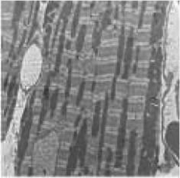
Transmission electron microscopic micrographs of left ventricular heart tissues from FVB and MT mice with or without DAHP treatment. (A): FVB; (B): FVB-DAHP; (C): MT; and (D): MT-DAHP. Myocardial tissues in A, C and D appear normal with myofibrils composed of regular and uninterrupted sarcomeres separated by continuous rows of normal mitochondria. In the myocardium from FVB-DAHP mice, irregularly shaped mitochondria and myelin figures are randomly distributed between highly disrupted myofibrils.