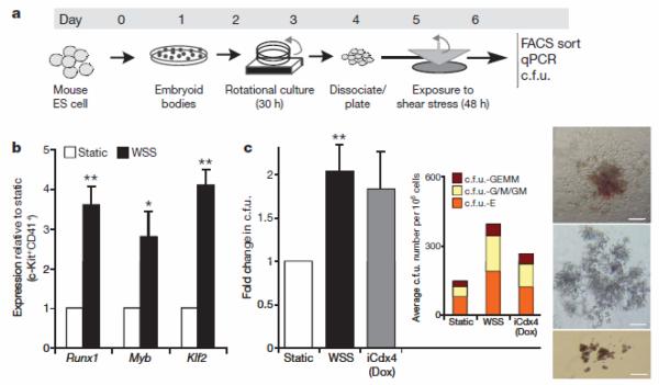
Figure 1. Shear stress induces haematopoietic commitment from ES-derived cells.
a, Experimental protocol used to induce haematopoietic differentiation from ES-derived cells in the presence of wall shear stress (WSS). ES cells are differentiated with the embryoid body method for 3.25 days, disaggregated and plated on gelatinized surfaces. Cell monolayers were exposed to shear stress and then collected on day 6 for further analysis b, Real-time Taqman PCR-based gene expression analysis in FACS-sorted CD41+c-Kit+ embryoid-body-derived haematopoietic precursors. Exposure to WSS induces upregulation of the haematopoietic markers Runx1 (P=0.01), Myb (P=0.03) and Klf2 (P=0.001), n=3. c, Methylcellulose haematopoietic c.f.u. assay. WSS increases the frequency of haematopoietic progenitors in complete M3434 methylcellulose. n=4, analysis of variance (ANOVA) P=0.01. Dox, doxycycline. Inset shows average distribution of haematopoietic colony types. c.f.u.-GEMM, c.f.u. granulocyte-erythroid-myeloid-megakaryocytes; c.f.u.-G/M/GM, c.f.u. granulocytes/macrophages/granulocyte-macrophages; c.f.u.-E, c.f.u. erythroid. Bar graphs represent average±s.e.m. Pictures show representative colonies: c.f.u.-GEMM (top), c.f.u.-G/M/GM (middle), c.f.u.-E (bottom). Scale bar, 200 μmm. *P<0.05, **P<0.01.

