Figure 5.
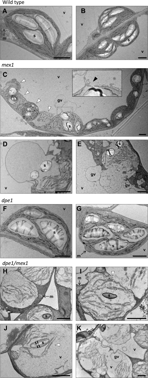
Chloroplast Ultrastructure in the Wild-Type, mex1, dpe1, and the dpe1/mex1 Double Mutant.
In each transmission electron micrograph, the bar = 2 μm. s, starch; v, vacuole; gv, globular vacuole; m, mitochondrion.
(A, B) Wild-type mesophyll cell chloroplasts.
(C) Multiple chloroplasts in a mesophyll cell from a mex1 mutant. Note the normal-looking chloroplasts on the right and the aberrant chloroplasts on the left (white arrowheads), which are misshapen, contain disorganized thylakoid membranes, and many plastoglobules. The swollen chloroplast in the center is associating with the adjacent globular vacuole. The inset shows an enlargement of the region of interaction (black arrowhead).
(D, E) Chloroplasts from mesophyll cells of the mex1 mutant that are either greatly swollen or that have fused with globular vacuoles. Many mesophyll cells of mex1 contained similar structures but none was seen in the wild-type.
(F, G) Chloroplasts from mesophyll cells of the dpe1 mutant. Note the enlarged starch granules.
(H) Swollen chloroplasts in three adjacent mesophyll cells of dpe1/mex1.
(I) Two fusing compartments, both containing chloroplast remnants, in a mesophyll cell of dpe1/mex1. Note the vesicles at the fusion site (white arrowhead).
(J) Remnants of a single chloroplast within a large vacuole in a mesophyll cell of dpe1/mex1. Note the absence of the chloroplast envelope (white arrowhead).
(K) A single cell of dpe1/mex1 containing multiple swollen chloroplasts and multiple vacuoles. One vacuole contains the remnants of three chloroplasts. Note also the numerous globular vacuoles. Cells with this appearance were common.

