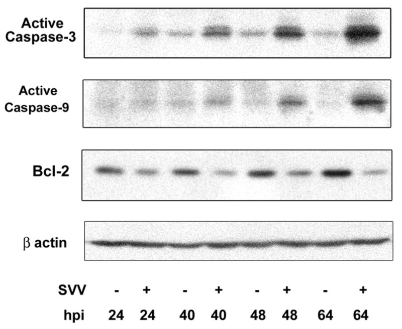
Figure 4. Intrinsic pathway of apoptosis in SVV-infected cells.

Uninfected (−) and SVV-infected (+) Vero cells were harvested at multiple times. Cell lysates were analyzed by Western blot analysis for the active forms of caspase-3 and caspase-9 and Bcl-2. Blots were reprobed for β-actin. Representative blots from four experiments are shown. Note the increased levels of caspases-3 and -9 and the decreased Bcl-2 expression in infected cells at 64 hr post-infection (hpi).
