
Figure 1.
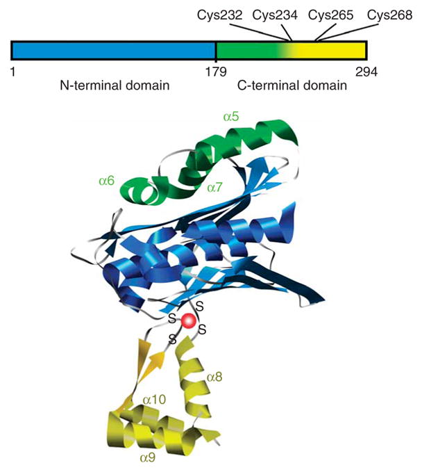
Domain structure of Hsp33. Linear representation (top) is color coded similarly to structural model of Hsp33 monomer (bottom), which is based on the crystal structure of the reduced Bacillus subtilis Hsp33 dimer (PDB 1VZY)10.
Official websites use .gov
A
.gov website belongs to an official
government organization in the United States.
Secure .gov websites use HTTPS
A lock (
) or https:// means you've safely
connected to the .gov website. Share sensitive
information only on official, secure websites.
Domain structure of Hsp33. Linear representation (top) is color coded similarly to structural model of Hsp33 monomer (bottom), which is based on the crystal structure of the reduced Bacillus subtilis Hsp33 dimer (PDB 1VZY)10.