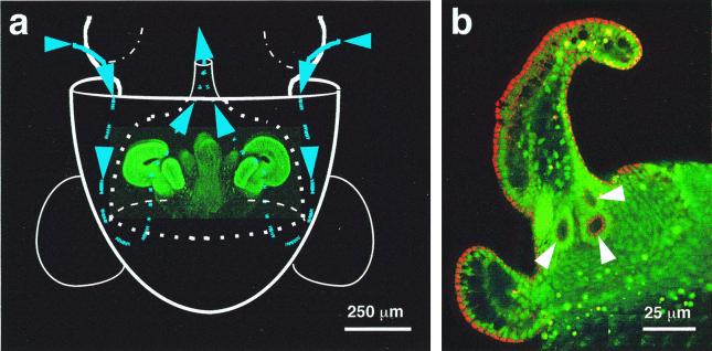
Figure 1.
The path of V. fischeri cells to the site of inoculation of the E. scolopes light organ. (a) Diagram illustrating an outline of the host's body (solid white lines), superimposed over a laser-scanning confocal micrograph (LSM) of the nascent light organ, indicating the relative size and position of the organ within the host's mantle cavity. The organ is circumscribed by the posterior portion of the excurrent funnel (dotted white lines). Ventilatory movements of the host draw ambient seawater (blue arrows and lines) containing V. fischeri cells into the mantle cavity. The water travels into the funnel where, before being vented back into the environment, it encounters complex ciliated fields (bright green) on the lateral surfaces of the organ. The fields entrain water into the vicinity of pores on the light organ surface. (b) Higher-magnification LSM of one side of a hatchling light organ, showing the location of the three pores (arrows) that lie at the base of the appendages of each ciliated field.

