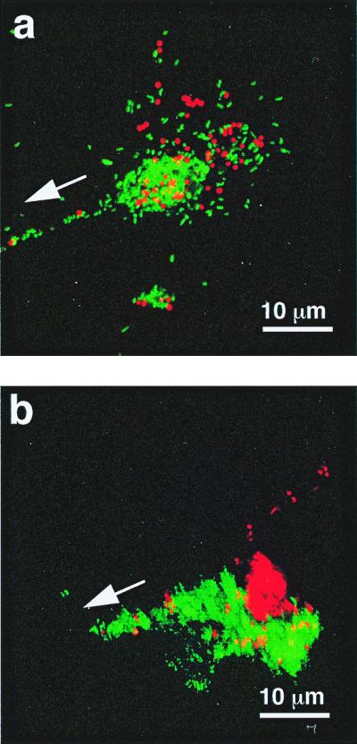
Figure 3.

The aggregation and segregation of polystyrene beads within the mucus-like secretions induced by the presence of V. fischeri cells. (a) Red-fluorescent polystyrene beads (Molecular Probes), 1 μm in diameter, were incubated with GFP-labeled V. fischeri and visualized by laser-scanning microscopy. After a 2- to 4-h incubation, bacteria and beads were found randomly distributed in aggregations. (b) Six hours after inoculation, the bacteria and beads had become segregated as the V. fischeri cells migrated in the direction of the pores (arrows).
