
Figure 3.
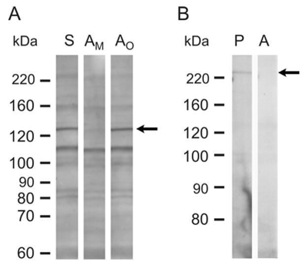
Western blots of soluble proteins from adult retinas immunostained with mMyo3B or mMyo3A. A. mMyo3B staining with total antiserum (S) and mMyo3B antiserum preincubated with Myo3B antigen (AM) or a His-tagged polypeptide with the sequence of the C-terminus of Limulus opsin1 antigen (AO). Antisera were used at a 1:500 dilution. The mMyo3B antiserum (S) and the antiserum preincubated with Limulus opsin1 (AO) immunostained the same multible protein bands in the retinal extract. On a replicate Western blot incubated in parallel with antiserum preincubated with Myo3B (AM) antigen a 130 kDa immunoreactive band (arrow) was selectively eliminated. B. mMyo3A staining with affinity purified antiserum (P) and affinity purified antiserum that had been preincubated with Myo3A antigen (A). Antisera were used at a 1:200 dilution. The immunopurified antiserum (P) stained a single band at 220 kDa which was not detected on blots incubated with antiserum that had been preincubated with Myo3A antigen. Molecular weight standards are shown on the left of each blot.