
Figure 8.
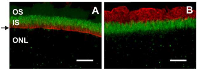
mMyo3A concentrates in the distal portion of the photoreceptor IS, does not distribute uniformly with F-actin and does not extend into OS. Fixed frozen sections were incubated without antigen recovery with affinity purified mMyo3A antibodies (undiluted) and A. fluorescently labeled phalloidin (1:200 dilution) or B. a monoclonal antibody directed against rhodopsin (1:200 dilution). Images were obtained as described in Fig. 6. A: mMyo3A-ir (green) is concentrated in the distal portion of the IS whereas F-actin, as visualized with fluorescently labeled phalloidin (red) is most abundant in the proximal portion of the IS and in the outer limiting membrane (arrow). B: mMyo3A (green) does not co-localize with rhodopsin (red) indicating that mMyo3A does not extend to the OS. Abbreviations as in Fig. 5. Bars=20 μm.