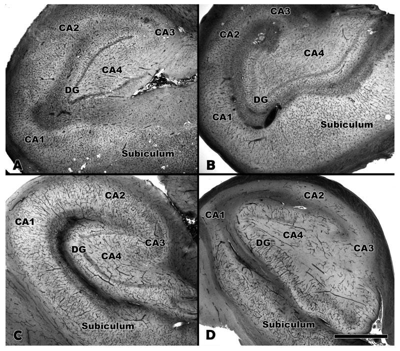
Figure 1.
Collagen type IV and alkaline phosphatase (AP) staining of hippocampi. (A, B) Total vasculature is labeled with antibody to collagen type IV. (C, D) Afferent vessels labeled by alkaline phosphatase histochemistry. Control hippocampus (A, C) and MTS hippocampus (B, D) are shown at 2× magnification. The density of afferent vessels (D) is markedly reduced in the MTS hippocampus compared to the control hippocampus (C). MTS, mesial temporal sclerosis; DG, dentate gyrus; CA, cornu Ammons. Bar = 1 mm.

