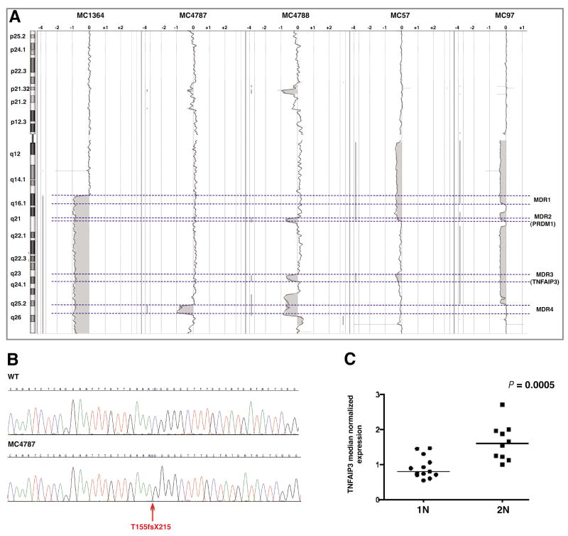
Figure 3.
TNFAIP3 abnormalities. A) Delineation of four minimal deleted regions (MDRs) on 6q based on aCGH data (dashed lines). PRDM1 and TNFAIP3 were localized in MDR2 and MDR3, respectively. B) Partial DNA sequences from a normal sample (top) and one with TNFAIP3 frame-shift deletion (red arrow; T155fsX215). The absence of the wild type allele indicates the homozygous status of the mutation. The position of TNFAIP3 mutation at protein level is based on NP_006281.1, which represent the accepted full length TNFAIP3 polypeptide. C) The TNFAIP3 transcript expression level was significantly lower in patients with one copy of the gene (1N) when compared with the group with two copies of TNFAIP3 (2N).

