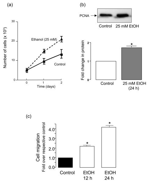
Figure 2. Ethanol stimulates HUVEC growth and migration.
HUVEC were treated with growth media in the absence (control) or presence of ethanol (25 mM) as described in Methods. (a) Cell counts of parallel triplicate wells were made on a daily basis. (b) Proliferating cell nuclear antigen (PCNA) protein expression in control and ethanol treated (25 mM, 24 h) endothelial cells; representative Western blot (top), cumulative densitometric data, n=3 (bottom). (c) Bar graph showing increased migration (scratch wound assay) by ethanol treated HUVEC at 12 and 24 h compared to control cells. *p<0.05 vs respective control.

