
Figure 3. Expression of Collagen 1 and Fibronectin in orthotopic tumor sections.
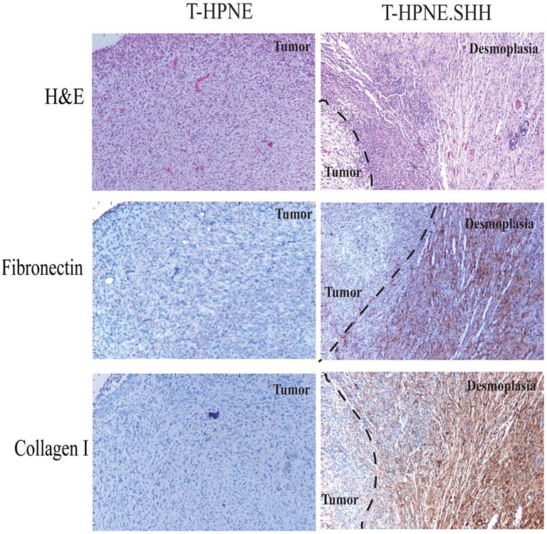

A. Representative immunohistochemical analysis of T-HPNE and T-HPNE.SHH tumor sections to determine if Fibronectin and Collagen 1 are expressed in either of the tumor sections. Areas of desmoplasia were identified using staining for SMA (described in Figures 1 and 2). Evidence that the SMA+ areas were expressing fibronectin and collagen 1 is indicated by brown staining in the areas of the tumor sections designated desmoplasia in the T-HPNE.SHH tumor sections. We did not observe positive staining in tumors derived from the T-HPNE cell line. H&E staining of the tumor sections is also shown.
B. Representative IHC staining for Collagen 1 and Fibronectin in the Capan-2 orthotopic tumors with no treatment, treatment with 4E11 or treatment with 5E1. There was a decrease in the intensity and amount of staining for both Collagen 1 and Fibronectin in the tumor sections treated with 5E1 when compared to the controls.