
Figure 2. PTEN loss confers growth in minimal media.
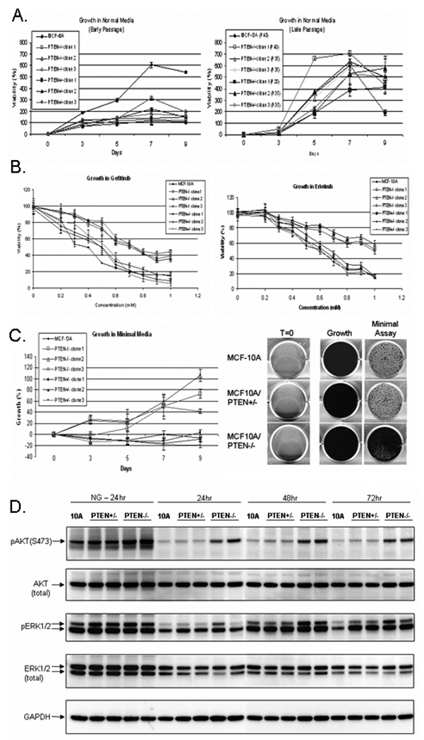
(A.) Early and late passage growth of MCF-10A cells, PTEN+/− clones, and PTEN−/− clones in normal MCF-10A growth media. Points, means of 2 independent experiments performed in quadruplicate; bars, SD. (B.) Cell growth of MCF-10A cells, PTEN+/− clones, and PTEN−/− clones after exposure to increasing doses of the EGFR antagonists Gefitinib and Erlotinib. Points, means of 3 independent experiments performed in quadruplicate; bars, SD. (C.) Cell growth in minimal media. Points, means of 2 independent experiments performed in quadruplicate; bars, SD. On day 6, MCF-10A cells, PTEN+/−, and PTEN−/− cells were fixed and stained with a solution of 10% PBS-buffered formalin and 0.25% crystal violet. Representative wells from the MCF-10A, PTEN+/−, and PTEN−/− cells are shown. (D.) MCF-10A, two PTEN+/−, and two PTEN−/− clones were plated in either normal growth media (NG) or minimal assay media and harvested at the indicated times by direct addition of RIPA lysis buffer.