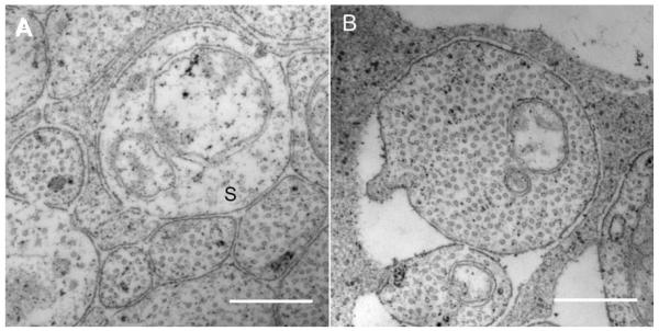
Figure 4.
High magnification images (28000X to 44000X) were used to measure RGC axon neurotubules and organelles. Sparse axons were rare in nerve fiber bundles except in the temporal macular region of the eye in both subjects. Axons that had a neurotubule density that was less than 25% percent of the average neurotubule density of the axon population were classified as sparse axons. (A) Sparse axons (S) appear pale and have fewer nerurotubules. Scale bar, 0.5 μm. (B) A RGC axon from nasal region of subject 57204. Scale bar, 0.5 μm.

