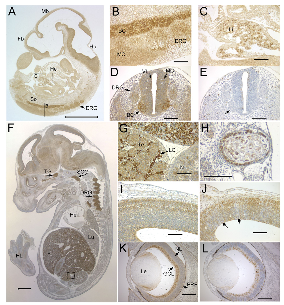
Figure 2. Developmental expression pattern of NSDHL in mouse embryos.
A. A saggital section from an E10.5 WT embryo immunostained for NSDHL showed the highest signal in the CNS and fetal liver. B. A higher magnification view of boxed area B in panel A showing intense staining of the basal column (BC) of the neural tube, with less signal in the mantle column (MC) and caudal dorsal root ganglia (DRG). C. A higher magnification view of boxed area C in panel A showing a subpopulation of strongly stained cells in the fetal liver (Li). D. A transverse section through the posterior neural tube illustrates the high level of NSDHL staining in the basal column of the neural tube relative to the mantle column and ventricular layer (VL). E. A section adjacent to that shown in panel D immunostained for phosphorylated histone H3, that marks mitotic cells, shows numerous dividing cells in the ventricular layer lining the neural tube, with no positive cells in the basal column, where the strongest NSDHL signal is seen (arrow). F. A saggital section from an E14.5 WT embryo immunostained for NSDHL shows intense staining in the liver, dorsal root ganglia, and trigeminal ganglion (TG). Signal in the brain is higher than in other tissues such as heart (He) and lung (Lu). G. A high magnification view of the boxed area from panel F, showing strong staining of the Leydig cells (LC) in the testis, comparable to the level in hepatocytes seen in the adjacent fetal liver. H. High magnification view of developing rib from section shown in panel F. NSDL staining is seen in cells of the condensing mesenchyme surrounding the chondrocytes of the rib. I. A high magnification view of the posterior neopallial cortex of the brain from the section shown in panel F. J. The same region of the brain shown in panel I from a Bpa1H E14.5 female embryo was immunostained for NSDHL. Note the patchy staining pattern, with radiating sectors of neurons showing no staining (arrows). K. A transverse section through the eye of a WT E15.5 embryo showing strong NSDHL staining in the ganglion cell layer (GCL). L. NSDHL staining of a Bpa1H E15.5 eye, showing mosaic expression in the GCL with radiating sectors of NSDHL positive and negative cells in progeny cells of the developing retina. Sections in panels B–L were lightly counterstained with hematoxylin. The size bars in panels A and F represent 1 mm. Size bars in all other panels denote 100 µm. Abbreviations: BC, basal column; DRG, dorsal root ganglion; Fb, forebrain; GCL, ganglion cell layer; Hb, hindbrain; He, heart; HL, hind limb; Ki, kidney; LC, Leydig cell; Le, lens; Lu, lung; Mb, midbrain; MC, mantle column; NL, neuroblast layer; PRE, pigmented retinal epithelium; VL, ventricular layer.

