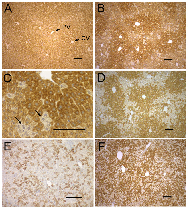
Figure 3. Immunostaining of NSDHL in WT and Bpa1H liver.
A. Adult (3 mo) WT liver shows strong staining thoughout the lobule, with slightly higher signal in the pericentral region. B. An adult (13 mo) Bpa1H liver with a large majority of NSDHL positive cells. C. A high magnification view of the liver shown in panel B showing small clusters of hepatocytes that did not stain for NSDHL. D. An adult (13 mo) Bpa1H liver with a relatively large number of NSDHL negative hepatocytes. Note the high proportion of NSDHL positive cells surrounding the central veins. NSDHL staining in the liver of a P6 Bpa1H female. F. NSDHL staining in the liver of a P25 Bpa1H female. Abbreviations: CV, central vein; PV, portal vein. Size bars represent 100 µm.

