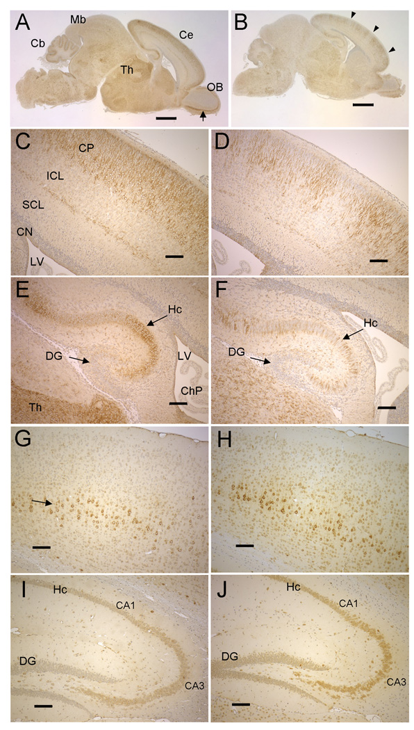
Figure 5. NSDHL in early postnatal and adult brains of WT and Bpa1H females.
A. NSDHL staining of a saggital section from a WT P2 brain. The arrow indicates the mitral layer of the olfactory bulb. B. NSDHL staining of a Bpa1H P2 brain. Arrowheads indicate radial sectors of low NSDHL signal in the cerebrum. C. A higher magnification of the cerebral cortex from the section shown in panel A shows a broad, even band of positively stained pyramidal neurons in the outer layer of the cortex. D. A higher magnification of the cerebral cortex from the Bpa1H brain section shown in panel B reveals regions of unstained cortical neurons that were not seen in the WT brain. E. A higher magnification of the hippocampus (Hc) from the WT brain shown in panel A. F. High magnification of the hippocampus from the Bpa1H brain shown in panel B, showing a mosaic pattern of NSDHL within the hippocampus. G. NSDHL staining in the cerebral cortex on a saggital section of a WT adult brain. The arrow indicates a layer of relatively large pyramidal neurons that showed the highest level of NSDHL signal in the adult cerebral cortex. H. NSDHL staining in the cerebral cortex of a Bpa1H adult brain. I. NSDHL staining in the hippocampus of a WT adult brain. J. NSDHL staining in the hippocampus of a Bpa1H adult brain. Abbreviations: Cb, cerebellum; Ce, cerebrum; ChP, choroid plexus; CN, cortical neurepithelium; CP, cortical plate; Cx, cerebral cortex; DG, dentate gyrus; Hc, hippocampus; Ht, hypothalamus; ICL, intermediate cortical layer; LV, lateral ventricle; OB, olfactory bulb; SCL, subventricular cortical layer; Th, thalamus. Size bars in panels A and B correspond to 1 mm. Size bars in panels C–J correspond to 100 µm.

